Scientists Discover Cell Behaviour Inside Healthy, Diseased Organs

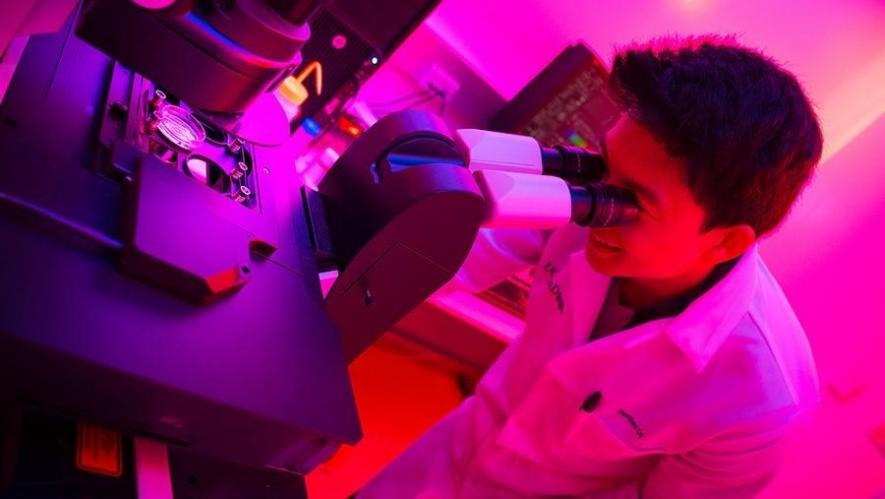
Image source: UC San Diego. Used for representation only.
Imagine a voyage through the layers of tissues in an organ, say the kidney, and you see how the cells are lined up and travel just like one sees roads, households and terrain travelling through a place.
This is not the plot of a science fiction story. Now, scientists can see cells behaving in their way inside an organ and the deviation in their behaviour when a disease strikes.
In a recent series of three research published in Nature, a group of scientists reported having mapped the kidney, intestine and placenta cells (the three research papers can be found here, here and here).
In their atlases, the scientists aimed to map the cells, how they direct the maternal blood supply in the placenta, how they transform in a diseased kidney and how the cells in the intestines organise themselves.
In an article by Heidi Ledford in Nature about the research, Michael Snyder, Stanford University geneticist and author of the intestine mapping study, said, “These cells organise themselves into neighbourhoods, towns and countries, and it affects their function.”
The three research represent an increasingly popular approach in studying body organs in healthy and diseased conditions. The research have hundreds of thousands of data depicting the gene activity and the resultant protein production in individual cells. These were subsequently mapped to the specific locations of the organs.
Technological advances have allowed researchers to track the gene activities inside a cell, meaning how genes express and how they govern cellular activity, etc. Scientists have previously created cell maps of other organs, especially the blood vessels of the brain and also of various types of tumours.
As time passed on, these technologies developed further. Now, researchers can track gene activity and locations of cells with more precision. Once, the discovery of types of cells inside a body was a great deal. But now scientists are not restricted merely in finding the types of cells but their activities in real time.
The latest Nature research are part of a consortium called the Human Biomolecular Atlas Program, having financial support from the National Institutes of Health, USA. This consortium aims to map out more cells and understand how they determine organs function. Remember, in a normal condition, cells having a particular behaviour determine an organ’s normal course of functioning, which gets deviated in diseases.
In the placenta study, the researchers studied the interface between the placenta and the uterus (the placenta develops in the uterus during pregnancy and provides oxygen and nutrients from the mother’s body to the baby).
With massive data of over 5 lakh cells and nearly 600 uterine arteries, the team attempted to understand it better. They could visualise how cells from the foetus invade the uterus and sort of remodel its blood vessels. This happens so that the blood vessels become larger and better at delivering nutrients as pregnancy progresses.
Michael Angelo, a Stanford University pathologist and an author of the placenta study, in a comment in Heidi Ledford’s article said, “They invade the arteries and replace the maternal cells, which is wild.”
Notably, the condition of pre-eclampsia distorts this process and then it becomes dangerous for both the foetus and the mother.
In the other study, scientists mapped the kidney cells’ behaviour in normal and diseased condition. Sanjay Jain, a Washington University School of Medicine pathologist and a co-author, seemed to be convinced with the team’s success when he said, “We were able to build pathways of how the cells might be travelling from healthy to injured, and the rest stops along the way.”
Having a complex structure, scientists have long struggled to develop kidney models that depict real kidney structures and function. The lack of model kidneys has made developing new drugs for kidney diseases harder.
The latest research comprises maps from 51 types of kidney cells with 28 cellular states representing injury or disease. Moreover, the researchers could also develop interactive 3D models of cells and microenvironments—all from 45 healthy people and 48 with kidney disease.
On the other hand, in the intestine mapping study, researchers studied cells taken from eight sites along the intestine. The map displayed layers of cells segregated into distinct groups, which they termed as neighbourhoods of cells having unique characteristics.
Scientists look forward to map other organs with their unique cells types and cellular behaviour to understand organ functioning both in normal and diseased conditions.
Get the latest reports & analysis with people's perspective on Protests, movements & deep analytical videos, discussions of the current affairs in your Telegram app. Subscribe to NewsClick's Telegram channel & get Real-Time updates on stories, as they get published on our website.


